Innovation features
1. Innovation backgrounds
We have researched for anti-solid cancers as follows.
Conventional chemosensitivity test determines the best anti-cancer drug and is often hires cell-based assay using human living cells. In cell-based assay, human cells are proliferated on a dish and incubated with anti-cancer drugs for a certain term, and a viable cell amount is counted. Many conventional chemosensitivity tests are available such as Succinate Dehydrogenase Inhibition test (SDI). But these are based on endpoint assay and require long culture duration.
Assays using two-dimensional (2D) growth of cells often necessitate time-consuming experimental processes (2 days) and rigorous laboratory conditions. Furthermore, the labeling might cause competitive interference with anti-cancer drugs and thereby degrade the reliability in the efficacy prediction of anti-cancer drugs. Moreover, 2D assays will not evaluate polypharmaceutical efficacy. The 2D assays are often hired by the reasons of easy to handle and of cost effectiveness. However, they will not provide the physiological concentration of drugs; that is the concentration level practically applied to a patient. This is caused by the cell growth environmental difference from the in vivo environment such as deficiency of extra cellular matrix. For example, the results obtained are 10 to 100 times higher against in vivo level concentration. Therefore, 2D assays are often used to focus on efficacy exits or not.
Optimized assays contain cells and organs keeping cell microenvironment, but they have issues such as maintaining the complicated culture system healthy. From these, the model reconstitution is performed on in vivo environment for in vivo-like cell-based experiment to achieve the evaluation of therapeutic potentials of anti-cancer drugs. For this, three-dimensional (3D) assays are hired, but necessitate time-consuming experimental procedure for about 2-4 weeks; the 3D assays cannot be automated easily. Especially in Collagen Gel Droplet embedded culture Drug Sensitivity Test (CD-DST), the obtained results are correlated to those from clinical tests of many solid cancers. However, labeling agents used sometimes cause low reliability of compound efficacy prediction similar to 2D assays.
New chemosensitivity tests such as Microphysiometer measure cellular statuses, namely extracellular fluxes of H+ (pH), O2, and drug, and cell morphology changes by impedance and current changes of living cells cocultivated with target compounds, sometimes using a semiconductor. The combination of integration of sensors and cell culture techniques including 3D method advances label-free cell-based technology for real-time and direct cell monitoring methods with drug flow. In case of using collagen, new chemosensitivity tests will give the concentration information at in vivo level. Furthermore, it can evaluate drug resistance and toxicity of a target compound.
However, measured traits are not keys for final efficacy evaluation and are dependent on a drug mode of action. Furthermore, sensing principals such as current and impedance cause artifacts (toxicity) on living cells. From these, many troubles happen in monitoring the essential cell signals to lead low compatibility with a standard chemosensitivity test and low reliability in polypharmacy. Moreover, new chemosensitivity test requires long cell culture with medium exchange (half of endpoint against a standard chemosensitivity test) causing large test errors and long decision duration (Referred paper 3 and Comparative paper 1-3). Using omics, markers that predict chemosensitivity have been studied. However, for example, the pharmacodynamic interactions of drugs, often enhancing the therapeutic potential of individual drugs, are still unpredictable in many cases by markers because a cancer patient might exhibit unconventional aspects of tumor development.
For these reasons, a rapid, in vivo-like label-free non-invasive cell-based assay is greatly anticipated for use in verifying the prediction of anti-cancer drug efficacy, both quantitatively and simply. The emerging assay reflects the in vivo anti-cancer drug response in patients, as evidenced by the close relation between the emerging assay and conventional chemosensitivity testing associated with in vivo drug response.
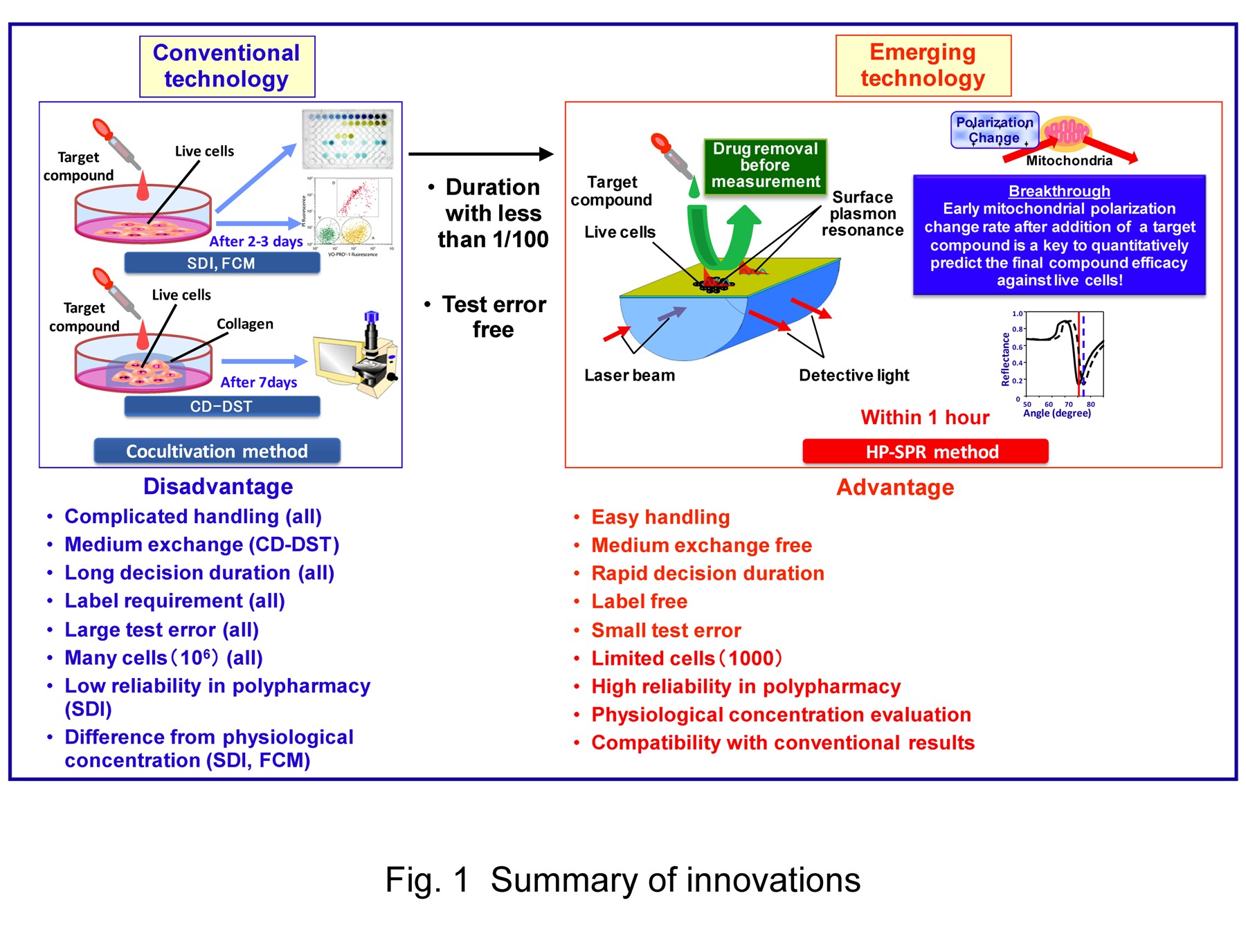
2. Innovation features (Fig. 1)
We developed our original device and method against the demand described above as follows.
(1) Prediction of final status (endpoint) without cell culture + Compatibility with conventional results
A surface plasmon resonance (SPR) sensor enables to detect the interactions between various molecules for the affinity and kinetics studies without the problems in a label-free and real-time system since SPR detects binding-induced refractive index changes of the dielectric on the metal surface. On the other hand, cell activity is often described as the dielectric polarization level. We hypothesized SPR might detect cell activity change by a short duration of culture with a medium. However, unknown detected signals of the SPR angle changes have to be explained by other offline methods such as a fluorescence dye. Furthermore, we needed a high precision SPR to detect the small change of dielectric polarization level of cells rapidly. First, we developed a new high precision SPR (HP-SPR) sensor (average fluctuation; 50 ndeg s-1) to solve the sensitivity issue.
Secondary, we established a new methodology for a rapid verification in apoptotic efficacy of drugs using 103 living solid cancer cells (pancreatic, liver, breast and small lung) attached onto a sensor chip. The HP-SPR measurement was performed with anti-cancer drugs such as Herceptin (Trastuzumab), doxorubicin, and paclitaxel. The time-course cell reaction was monitored as the HP-SPR angle change rate for ca.10 min during 1h cell culture with one or combined drugs. It was significantly related to cell viability counted after 48h of 2D cell culture.
We have succeeded the development of new original rapid and reliable screening system regardless to compound mode of action with label-free manner. This included high reliability in polypharmacy without using indirect and statistic methods such as genomics. We have achieved toxicity evaluation using normal (fibroblast) cells.
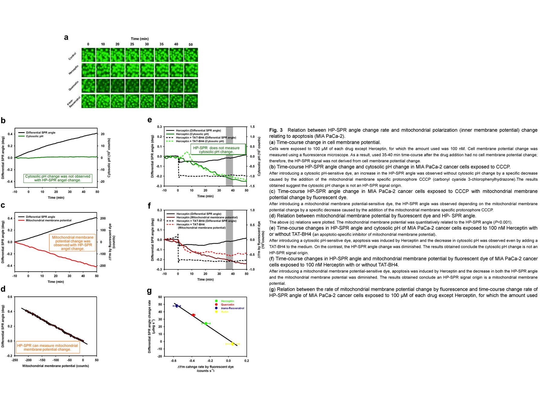
HP-SPR angle change origin was examined using a fluorescence microscope equipped over an SPR sensor for simultaneous detection.
The detected HP-SPR signal was derived from the decrease in mitochondrial membrane potential (Fig. 3). Because our HP-SPR system performs efficacy prediction at endpoint without cell culture, its principle is different from conventional chemosensitivity tests and from new chemosensitivity tests such as Microphysiometer (Patent papers 1-3, Referred papers 1-3)。
We have found that early mitochondrial polarization change rate after addition of a target compound is a key to quantitatively predict the final compound efficacy against living cells. Here, mitochondria is reported as a qualitatively concern to cell viability. We validated its quantitative concern. And we proved the HP-SPR signal related to conventional chemosensitivity test at non in vivo level for the efficacy and toxicity.
The features of our systems are summarized as follows.
a) HP-SPR
High precision for our system was achieved by high precision prism, beam molding, temperature control, low vibration, algorithm and so on, and reached 50 ndeg s-1 average fluctuation. The cells self-attached onto a gold surface sensor chip allow selective measurement of mitochondria polarization status within cells at high sensitivity (dielectric constant of 100 n/s order) as the average of whole cells quantitatively. Our system does not detect pH change within cells.
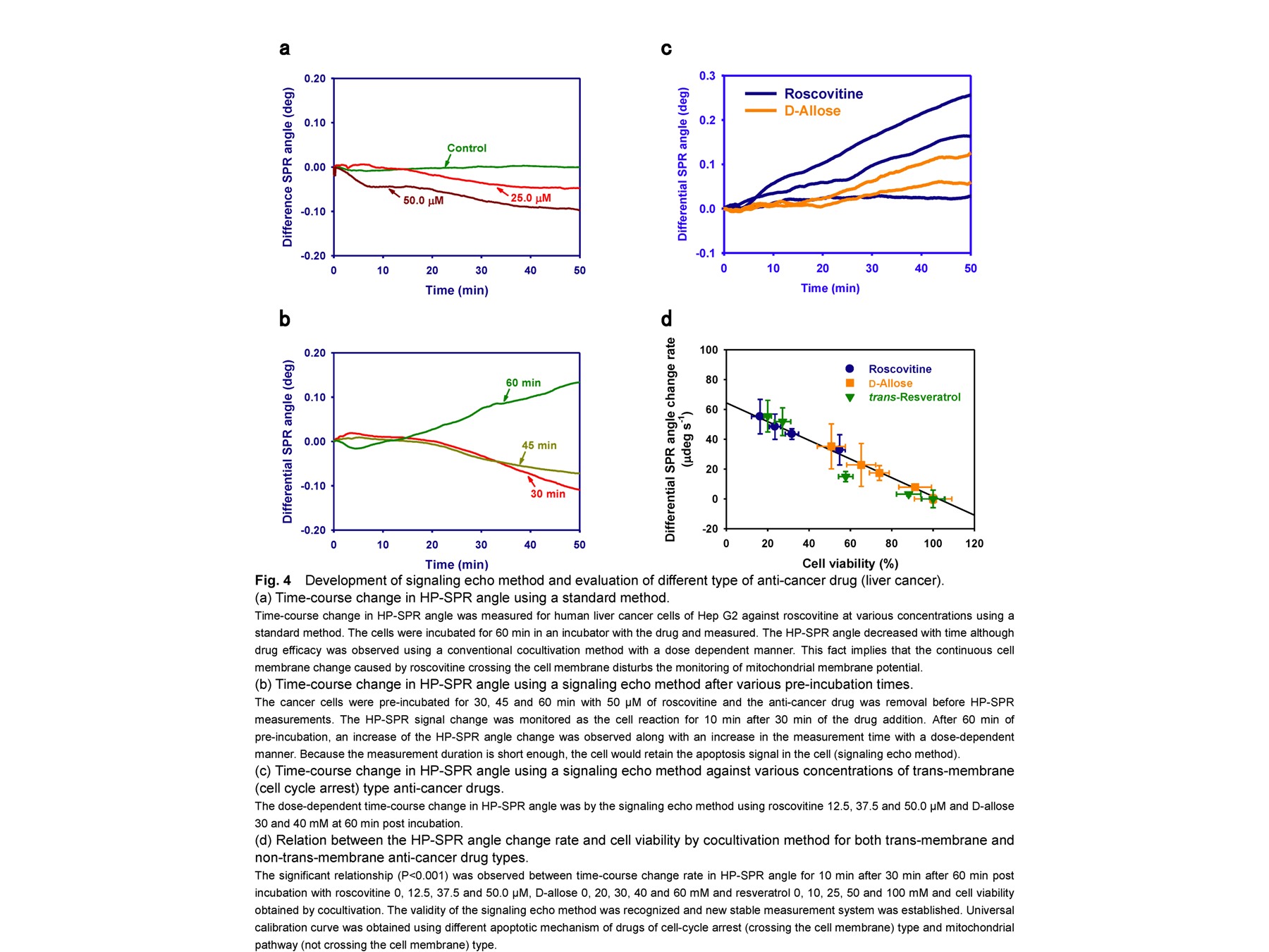
b) Signaling echo method (Fig. 4)
For trans-membrane anti-cancer drugs to be effective, continuous potential changes arising from the crossing of the membrane by the drug of interest prevent mitochondrial polarization status monitoring. Herein, we developed on a novel signaling echo method that avoids this disturbance; the cancer cells are incubated with a specific anti-cancer drug for 1h, with subsequent removal of the drug before HP-SPR measurements. This is based on the short duration of HP-SPR measurement and on the signals of mitochondrial polarization changes retained in the cell, even after drug removal. This is advanced in selective mitochondrial polarization status monitoring.
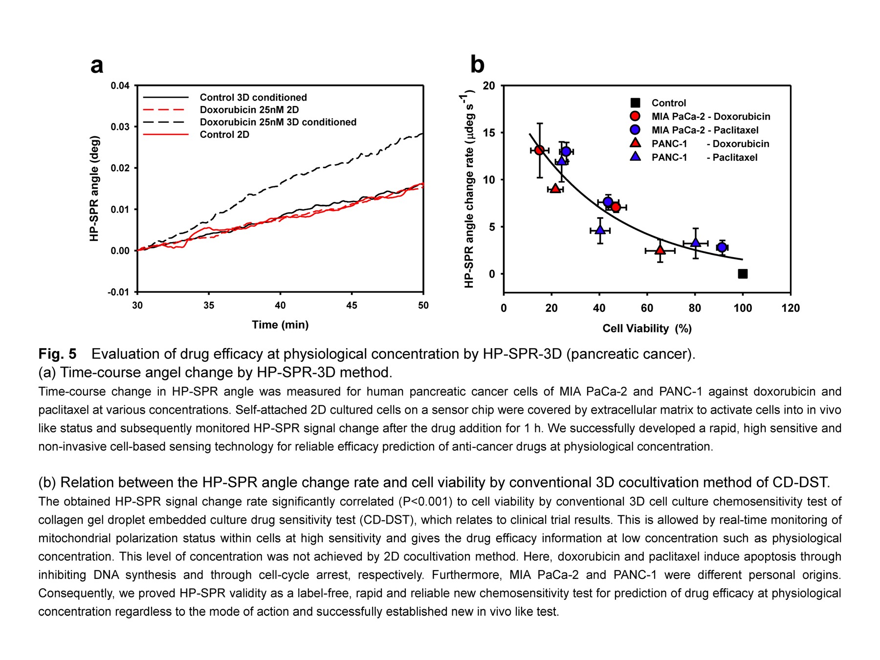
(2) Monitor of in vivo-like cell status using 2D cells by low energy scan to avoid triggering toxicity + Reproduction of in vivo-like cell status using 2D cultured cells (Fig. 5)
We succeeded to modify HP-SPR method for evaluation of anti-cancer drugs at physiological concentration, using pancreatic cancer cells (HP-SPR-3D). To achieve this, self-attached 2D cultured cells on a sensor chip were covered by collagen for several hours and subsequently monitored HP-SPR signal change after the drug addition for 1h. The obtained HP-SPR signal change rate correlated to cell viability by conventional 3D cell culture chemosensitivity test of Collagen Droplet embedded culture Drug Sensitivity Test (CD-DST) and proved its validity as a label-free, rapid and reliable new chemosensitivity test for prediction of drug efficacy at physiological concentrations regardless to the mode of action (Patent paper 3). We proved that living cells were activated into 3D cell culture status like in vivo by covering the cells with collagen for a few hours before the cell division.
(3) Prediction of next or final differentiation status of stem cell at the initiation non-invasively
Based on the in vivo-like test technology described above, we enable the prediction of next or final differentiation status of stem cell at the initiation (Patent paper 2). The monitoring of mitochondrial polarization status including distribution within cells is revealed as a key to predict the destination of cells by differentiation.
Our technology does not require isolation of cells from a culture plate and tests all cells used to avoid contamination and to achieve high purity. Furthermore, high safety is kept because future cancerous cells are screened by our technology. Additionally, high activity cells are screened by monitoring mitochondrial activity.
3. Competing products and similar products
For solid cancers, competing products to our system are new chemosensitivity test device such as Microphysiometer. The measured traits are extracellular fluxes of H+ (pH), O2, and drug, and cell morphology changes by impedance and current changes of living cells cocultivated with target compounds, sometimes using a semiconductor. The combination of integration of sensors and cell culture techniques including 3D method advances label-free cell-based technology for real-time and direct cell monitoring methods with drug flow. In case of using collagen, new chemosensitivity tests will give the concentration information at in vivo level. Furthermore, it can evaluate drug resistance and toxicity of a target compound.
However, measured traits are not keys for final efficacy evaluation and are dependent on a drug mode of action. Furthermore, sensing principals such as current and impedance cause artifacts (toxicity) on living cells. From these, many troubles happen in monitoring the essential cell signals to lead low compatibility with a standard chemosensitivity test and low reliability in polypharmacy. Moreover, new chemosensitivity test requires long cell culture with medium exchange (half of endpoint against a standard chemosensitivity test) causing large test errors and long decision duration. No competing products exist. As far as SPR, many instruments exist but not for monitoring mitochondria of living cells and do not give comparable results to conventional chemosensitivity test such as 3D cell culture.
For floating cancers such as acute lymphocytic leukemia, a rapid chemosensitivity test device is not found.
Furthermore, our system does not require cell culture and is very effective for new drug screening especially in using stem cell disease model necessitating long term cell culture causing difficulty to keep the healthy cell condition. From this point, our system is very unique.
On the other hand, we enable non-invasive prediction of next or final differentiation status of stem cell at the initiation using the same HP-SPR method.
Consequently, there is no competing products and no similar products to ours.
Patent paper
- Ona, T. and Kosaihira, A. Monitoring of intercellular mitochondrial polarization. WO2007/069692 A1 (2007); CN 200680052043.8 (2008); EP 1 961 824 A1 (2008); US 2011/0003321 A1 (2011); JP2012-93369A (2012).
- Nomura, M., Matsubara, E., Ona, T. Determination system, selection system, determination method, cell production method, program, and recording medium. JP2009-122016A (2009).
- Ona, T., Method for activating two-dimensional cultured cells similarly to three-dimensional culture or in vivo, and use thereof. WO 2013/039112 A1 (2013).
Referred paper
- Kosaihira, A. and Ona, T. Rapid and quantitative method for evaluating the personal therapeutic potential of cancer drugs. Anal Bioanal Chem, 391:1889 (2008).
- Nishijima, H., Kosaihira, A., Shibata, J. and Ona, T. Development of signaling echo method for cell-based quantitative efficacy evaluation of anti-cancer drugs in apoptosis without drug presence using high-precision surface plasmon resonance sensing. Anal Sci, 26:529 (2010).
- Ona, T. and Shibata, J. Advanced dynamic monitoring of cellular status using label-free and non-invasive cell-based sensing technology for the prediction of anti-cancer drug efficacy. Anal Bioanal Chem, 398:2505 (2010).
Comparative paper
- Hatok, J., Babusikova, E., Matakova, T., Mistuna, D., Dobrota, D. and Racay, P. In vitro assays for the evaluation of drug resistance in tumor cells. Clin Exp Med, 9:1 (2009).
- Mestres, P. and Morguet, A. The Bionas technology for anticancer drug screening. Exp Opin Drug Discov, 4:785 (2009).
- Wlodkowic, D. and Cooper, J.M. Tumors on chips: oncology meets microfluidics. Cur Opin Chem Biol, 14:556 (2010).